Bruker NanoWizard 4 XP Bioscience AFM
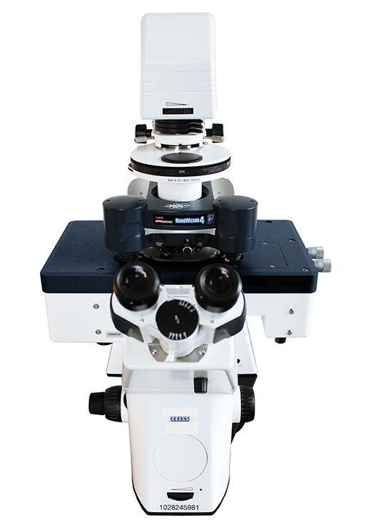
Bruker NanoWizard 4 XP Bioscience AFM
High-resolution imaging with extreme performance

The NanoWizard 4 XP BioScience atomic force microscope combines atomic resolution and fast scanning with rates of up to 150 lines/sec and a large scan range of 100µm in one system. It is designed to provide highest mechanical and thermal stability on inverted optical microscopes during long term experiments on samples ranging from single molecules to living cells and tissues.
Features:
- PeakForce Tapping® for easy imaging
- Fast Scanning option with up to 150 lines/sec
- NestedScanner Technology for high-speed imaging of surface structures up to 16.5µm with outstanding resolution and stability
- New tiling functionality for automated mapping of large sample areas
- V7 Software with revolutionary new workflow-based user interface
- DirectOverlay™ 2 software for perfect integration and data correlation with advanced fluorescence microscopy platforms
- Vortis™ 2 controller for high-speed signal processing and lowest noise levels
Automated mapping of large sample areas with new tiling functionality
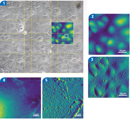 The HybridStage™ or Motorised Precision Stage transforms experiments by enabling direct access to a large sample area, with automated, motorised movement to selected positions, grids and mapping regions.
Begin with the DirectOverlay 2 optical calibration, and then select a region for optical tiling up to millimetres in size.
Precise motor movements automatically bring the whole sample into view, making it easy to select regions and features for further investigation. A single click navigates from point to point or MultiScan experiments automate a sequence of measurements at selected points.
The images show living Vero cells in cell culture medium at 37°C in the PetriDishHeater™ . [2] - [5] Optical tiling with 5×6 phase contrast images covering a 630µm×450µm region. - Zoom into region scanned with AFM showing 100μm×100μm scan (height range 5μm) and inset 15μm×15μm (height range 2μm) scan topography images using PeakForce Tapping. The feedback correction signal images highlight the surface membrane features, particularly in the zoomed image. Microvilli dominate the center of the cell, with membrane ruffles at the cell boundary.
For further information please contact us or download the datasheet.
