AX AXR Confocal Microscope

AX AXR Confocal Microscope
Nikon Corporation (Nikon) is pleased to announce the release of the next generation confocal microscope series, AX and AX R. This new confocal series features a completely re-designed scan head with 8K x 8K resolution*1, ultra-high speed resonant scanning, and world�s largest*2 25 mm field of view*3. Combined with a new user-friendly interface and advanced AI-based tools, the AX/AX R confocal series is designed to enable users to acquire data faster, with an unprecedented level of detail and ease.
1. Capture more data than ever before with 8K x 8K pixel density and a 25 mm field of view
The AX and AX R enables 8192 x 8192 pixel, high-resolution images to be obtained with the world�s largest field of view (25 mm). Now, ultrafine details can be easily captured even for large specimens. This feature is available for both inverted*4 and upright*5 microscope configurations, thereby supporting a wide range of research applications and fields.
In addition, with the AX R�s high-speed resonant scanner users can acquire up to 720 frames per second at 2048 x 16 pixels. High-speed resonant scanning not only enables dynamic events to be easily captured but also reduces the amount of time required to image large, fixed specimens.
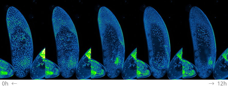
Time-lapse Z series maximum intensity projection images of a developing Drosophila embryo expressing PLC-PH::GFP (PIP2) acquired every 10 minutes for 12 hours with 2K x 1K pixels (with 25X silicone immersion objective), courtesy of Yang Hong Laboratory, Department of Cell Biology, University of Pittsburgh in collaboration with the Center for Biological Imaging. With high-speed scanning, it is possible to minimise photobleaching of the specimen even with long-term imaging.
2. Improved specimen viability for longer time-lapse imaging
The AX/AX R features detector units that are twice as sensitive as conventional models*6, with a ~30% reduction in dark current noise*7. With increased sensitivity and reduced noise, lower illumination power can be used for imaging even dim specimens, minimizing photobleaching and phototoxicity. Combined with high-speed resonant scanning which further decreases exposure time, specimen viability is greatly extended, enabling extreme, long-term time-lapse imaging.
3. A simplified, AI-driven user experience
Nikon�s NIS-Elements C imaging software allows users to easily customize the layout and experiment workflow to suit their needs. The new Autosignal.ai tool simplifies confocal acquisition by automatically determining the optimal illumination and detection settings while Denoise.ai removes shot noise from confocal images to enable clearer images with shorter exposure times. AI-based tools for post-acquisition image processing such as Segment.ai makes segmentation of complex structures more efficient and reliable.
Graphical programming tools for both acquisition and analysis provide further flexibility for the user. The optional JOBS module enables complex, non-linear experiments with multiple paths and dimensions to be created with ease, including conditional workflows where subsequent acquisition parameters are based on real-time analysis results.
For further information please contact us or read more.
*1 Galvano scanner
*2 Among point scanners available on the market as of 287 Apirl 2021, according to research conducted by Nikon
*3 The diameter of the observable image in a microscope
*4 ECLIPSE Ti2-E inverted research microscope
*5 ECLIPSE Ni-E motorised upright research microscope
*6 In cases where multi-alkali PMT is selected
*7 Noise generated by the heat of the sensor itself during long exposures
